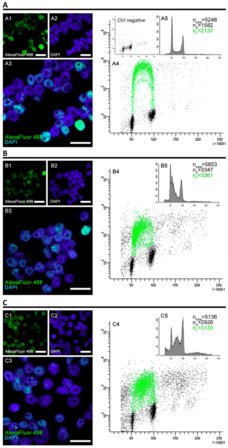
Figure 2.
The analysis of fluorescence levels and distribution of replicating clusters after 32 h of cell incubation. The cells were incubated in 100 µM EdU water solution for 0.5 h before the end of the 32-h experimental period and at the end of the incubation, the flow cytometry was performed. (A) Positive control, (B) negative control (induction of replicative stress), (C) PCC induction. The cells were stained with 4′,6-diamidino-2-phenylindole (DAPI) and antibodies conjugated with AlexaFluor 488. The binary images (A4,B4,C4) were prepared based on the thresholded originals. All the photographs are of the cells sorted by the flow cytometer’s sorting unit and include only the S-phase fraction. The scale bars are equal to 10 µm.

