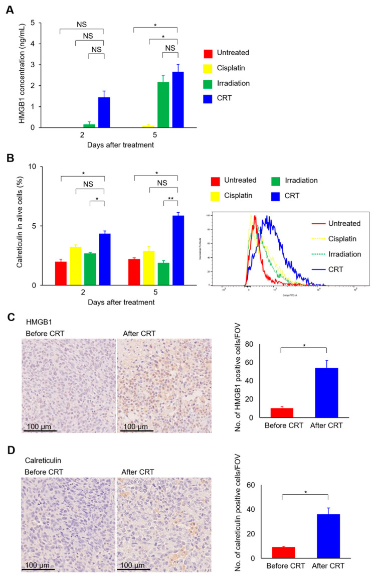
Figure 4.
The combination of cisplatin and irradiation increases HMGB1 protein secretion and cell surface expression of calreticulin protein in MB49 cells. (A) MB49 cells are treated with cisplatin at 0.6 mg/L and/or irradiated with a single fraction of 10 Gy. Medium is collected two and five days after treatment, and HMGB1 protein expression levels in each medium are examined in duplicate by ELISA (n = 3/group). (B) MB49 cells are treated with cisplatin at 0.6 mg/L and/or irradiated with a single fraction of 10 Gy. Cells are collected, and cell surface calreticulin protein expression levels are examined by flow cytometry (n = 3/group). (C,D) Seven days after inoculation MB49 cells in the left hindlimb, mice are treated with cisplatin at 3 mg/kg and/or irradiation with a single fraction of 10 Gy. Tumor sections before and after CRT (n = 3/group) are stained for HMGB1 (C) and calreticulin (D). Left, representative images; right, comparison of the mean (± standard error of the mean [SEM]) number of HMGB1- or calreticulin-positive cells counted in five fields of view (FOV). All data are shown as the mean ± SEM. Asterisks indicate p values comparing two groups, as indicated in the figure. NS, not significant; * p < 0.05; ** p < 0.01.

