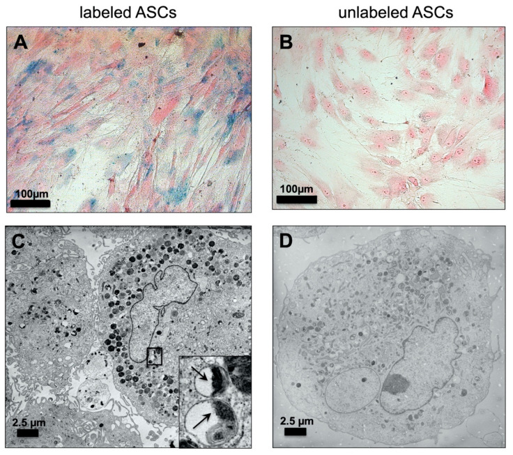
Figure 1.
Detection of VSOP-labeled hASCs: Prussian Blue staining of human adipose tissue-derived stromal cells (hASCs) labeled with 1.5 mM citrate-coated very small superparamagnetic iron oxide particles (VSOPs): (A) Intracellular blue spots indicate the uptake of iron oxide particles into the hASCs. (B) No blue staining was detected in unlabeled controls. Magnification ×200; scale bars represent 100 µm. Transmission electron microscopy (TEM) image analysis: (C) Intracellular distribution of vesicles containing VSOPs in hASCs after labeling with 1.5 mM VSOPs. The insert shows individual vesicles at higher magnification with intravesicular VSOPs highlighted by black arrows. (D) Within the unlabeled hASCs, no intracellular deposition of VSOPs was detected. Scale bars represent 2.5 µm.

