. 2021 Jan 22;10:e62387. doi: 10.7554/eLife.62387

Figure 3. Deletion of Runx2 in cranial neural crest (CNC)-derived cells leads to craniofacial defects in the soft palate.

(A, B) Intraoral views of palates from control and Osr2-Cre;Runx2fl/fl mice at newborn stage (P0). Arrow indicates the cleft in the posterior part of the soft palate. (C–F) Sagittal (C–D) and coronal (E–F) views of microCT scans of newborn control and Osr2-Cre;Runx2fl/fl mice (N = 3). Red arrows indicate the normal soft palate, and asterisks indicate the cleft in the posterior part of soft palate. (G–P) H and E staining of soft palate coronal sections from control and Osr2-Cre;Runx2fl/fl mice at P0 (N = 5). Yellow dashed lines outline the soft palate muscles. Black and red arrows in H and M show the pterygoid plate and tensor veli palatini (TVP) defects, respectively, of Osr2-Cre;Runx2fl/fl mice. Asterisks in N indicate the cleft soft palate in the levator veli palatini (LVP) region of Osr2-Cre;Runx2fl/fl mice. Boxed areas in G, I, L, and N are enlarged in H, J, M, and O, respectively. Boxed areas in J and O are enlarged in K and P, respectively. Scale bars in C-D and E-F indicate 0.5 mm and 0.9 mm, respectively. Scale bar in G indicates 400 µm for G, I, L, and N. Scale bar in H indicates 100 µm for H, J, M, and O. Yellow arrowheads in P indicate the centralized nuclei in mutant muscle cells.

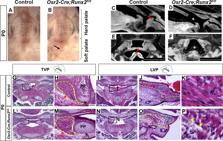
Figure 3.

Figure 3—figure supplement 1. Osr2-Cre specifically deletes Runx2 in the soft palate region.

Figure 3—figure supplement 1.

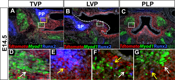
(A–G) Immunostaining of Runx2 and RNAscope in situ hybridization of tdTomato with myogenic marker Myod1 on coronal sections of tensor veli palatini (TVP), levator veli palatini (LVP), and palatopharyngeus (PLP) regions at E14.5. White dashed line outlines the palatal shelf in B. White and yellow boxes in A are enlarged in D and E. White boxes in B and C are enlarged in F and G, respectively. White arrows show the tdTomato+ cells surround all soft palatal muscles in Osr2-Cre;tdTomato mice in D, F, and G. Yellow arrows show colocalization of Runx2 and tdTomato in E, F, and G. Scale bar in A indicates 100 µm in A-C. Scale bar in D indicates 50 µm in D-G.
Figure 3—figure supplement 2. Deletion of Runx2 in cranial neural crest (CNC)-derived cells gives rise to hard tissue defects in Osr2-Cre;Runx2fl/fl mice.

Figure 3—figure supplement 2.

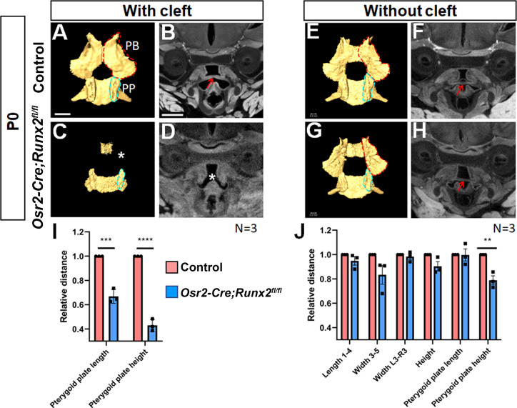
(A, C, E, G) Isolated palatine bones and sphenoid bones from control (A,E) and Osr2-Cre;Runx2fl/fl (C, G) mice. (B, D, F, H) Coronal views of soft tissue microCT scans of newborn control (B,F) and Osr2-Cre;Runx2fl/fl mice (D,H). (I): Quantification of the size (length and height) of the pterygoid plate from control (red bars) and Osr2-Cre;Runx2fl/fl (blue bars) mice. ***p<0.001; ****p<0.0001. (J): Quantification of the size (length, width and height) of the palatine bone and the size (length and height) of the pterygoid plate from control (red bars) and Osr2-Cre;Runx2fl/fl (blue bars) mice. **p<0.01. PB: Palatine bone; PP: Pterygoid plate. Red dashed lines outline the palatine bone. Blue dashed lines outline the pterygoid plate. White asterisk indicates the missing palatine bone or cleft. Red arrows indicate the normal soft palate. Scale bars in A and B indicate 0.6 mm for A, C, E, G and B, D, F, H, respectively.
Figure 3—figure supplement 2—source data 1. Source data for Figure 3—figure supplement 2I.
Figure 3—figure supplement 2—source data 2. Source data for Figure 3—figure supplement 2J.
Figure 3—figure supplement 3. Deletion of Runx2 in cranial neural crest (CNC)-derived cells gives rise to soft palate defects in Osr2-Cre;Runx2fl/fl mice.

Figure 3—figure supplement 3.

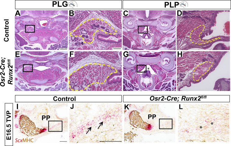
(A–H) H and E staining of soft palate coronal sections from control and Osr2-Cre;Runx2fl/fl mice at P0. Yellow dashed lines outline the soft palatal muscles. Asterisks in G indicate the cleft soft palate in the PLP region of Osr2-Cre;Runx2fl/fl mice. Boxed areas in A, C, E, and G are enlarged in B, D, F, and H, respectively. (I–L) RNAscope in situ hybridization of Scx and immunostaining of MHC in the tensor veli palatini (TVP) regions of coronal sections of E16.5 control and Osr2-Cre;Runx2fl/fl mice. Asterisks indicate the altered tendon structure in Osr2-Cre;Runx2fl/fl mice. Scale bar in A indicates 400 µm for A, C, E, G. Scale bar in B indicates 100 µm for B, D, F, H. Scale bars in I and J indicate 100 µm for I, K and J, L, respectively.
Figure 3—figure supplement 4. Deletion of Runx2 in cranial neural crest (CNC)-derived cells gives rise to aponeurosis defects in Osr2-Cre;Runx2fl/fl mice.

Figure 3—figure supplement 4.

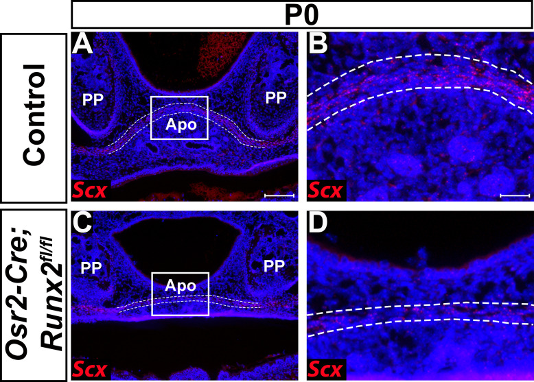
(A–D) RNAscope in situ hybridization of Scx of coronal sections in the tensor veli palatini (TVP) region of P0 control and Osr2-Cre;Runx2fl/fl mice. Scale bar in A indicates 100 µm for A and C. Scale bar in B indicates 25 µm for B and D.