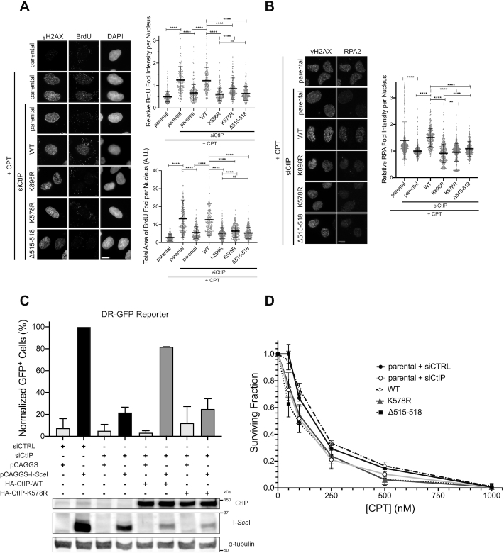
Figure 7.
Cells expressing K578R mutant CtIP show defects in DNA end resection and homologous recombination. (A) Left panel: parental U-2 OS cells or U-2 OS stably expressing the indicated siRNA-resistant GFP-CtIP constructs were transfected with siRNA to CtIP (or not) and treated with 1 μM CPT for 1 h (or not), then processed for IF staining. Cells were cultured in BrdU-containing media prior to treatment with CPT. Cells sensitive to CPT (seen by the induction of γH2AX foci, with the exception of the untreated condition) were quantified for BrdU foci (right panels). IF micrographs are representative of three independent experiments; 249–299 cells per condition from two independent experiments (total foci intensity) and 299–340 cells per condition from three independent experiments (total foci area) were quantified. (B) Similar to (A) but without BrdU in the culture media, and IF staining for RPA2 instead of BrdU. Micrographs are representative of four independent experiments; 467–748 cells per condition from three to four independent experiments were quantified for total RPA2 foci intensity. Asterisks depict statistically significant differences as determined by a two-tailed, unpaired, non-parametric Student's t-test (Mann–Whitney): ns (not significant), ** (P< 0.01), **** (P< 0.0001). (C) DR-GFP homologous recombination reporter assay in U-2 OS stably expressing the DR-GFP cassette. The cells were transfected with non-targeting (siCTRL) or CtIP-targeting (siCtIP) siRNA. 24 h later, they were transfected with the same siRNAs, and either pCAGGS empty vector or pCAGGS-I-SceI, in combination with siRNA-resistant constructs encoding HA-CtIP-WT or -K578R or not. GFP+ cells were assessed via flow cytometry 24 h later. Data presented are the means of two independent experiments (top panel). An immunoblot confirming the transfection combinations for one experiment is presented beneath (bottom panel). (D) Clonogenic survival assay of parental U-2 OS transfected with siCTRL or siCtIP, or U-2 OS cell lines stably expressing GFP-CtIP-WT, -K578R or Δ515–518 and transfected with siCtIP. Cells were treated with CPT at the indicated concentrations for 1 h, and colonies were allowed to form over ∼10 days. Survival data is presented as mean ± standard deviation from three independent experiments.

