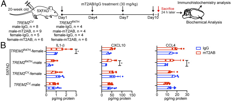
Fig. 7.
Sustained acute treatment with mT2AB affects microglial responses to pathology differently. (A) Schematic diagram of mT2AB treatment in TREM2CV-5XFAD or TREM2R47H-5XFAD mice. The 20-wk-old mice were injected intraperitoneally with murine mT2AB at 30 mg/kg every 3 d for 10 d. Littermates were administered the same concentration of control mIgG1 antibody. Mice were killed 24 h after the last antibody injection and brains were harvested for immunohistochemistry and biochemical analysis. (B) Quantification of cytokines and chemokines, such as IL-1β, CXCL10, and CCL4 in the cortex lysates among different treatment groups. *P < 0.05; ***P < 0.001; ****P < 0.0001 by two-way ANOVA with Sidak’s multiple comparisons test. Data are shown as mean ± SD. TREM2CV-5XFAD, male, mIgG1, n = 8; TREM2CV-5XFAD, male, mT2AB, n = 9; TREM2CV-5XFAD, female, mIgG1, n = 5; TREM2CV-5XFAD, female, mT2AB, n = 6; TREM2R47H-5XFAD, male, mIgG1, n = 4; TREM2R47H-5XFAD, male, mT2AB, n = 4; TREM2R47H-5XFAD, female, mIgG1, n = 4; TREM2R47H-5XFAD, female, mT2AB, n = 6.

