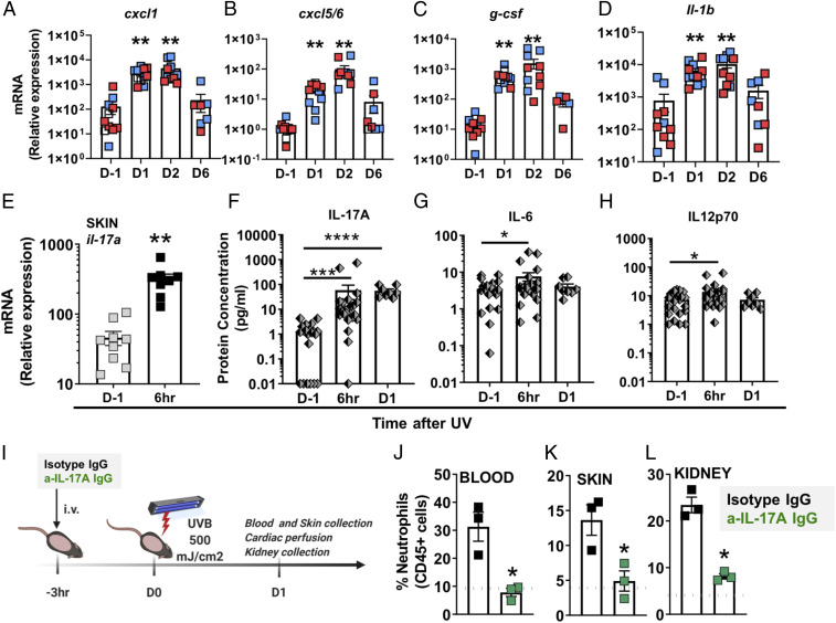
Fig. 3.
Skin exposure to UV light triggers IL-17A production, which is required for neutrophil recruitment. (A–D) Relative mRNA expression in skin obtained from female (red) and male (blue) mice was quantified by qPCR using the primers listed in SI Appendix, Table S1 and normalized to 18S transcript levels. The bars represent mean relative expression ± SEM for all samples combined. (E) Il-17A gene expression in the skin 6 h after UV exposure was quantified as in A–D. (F–G) Multiplex analysis of the protein concentration in plasma samples collected 6 h or one day (D1) after UV (F) IL-17A, (G) IL-6, and (H) IL-12p70. Significant changes in gene expression and plasma protein levels at different time points after exposure to UV light were determined relative to those of nonirradiated controls (D-1) using one-way ANOVA with Tukey’s post hoc (A–D, n = 8 to 10; F–H, n = 8 to 25 mice per group; mean ± SEM, *P < 0.05, **P < 0.01, ***P < 0.001). (I) Shaved B6 mice were treated intravenously (i.v.) with 100 µg anti–IL-17A IgG (green) or isotype control IgG (black) 3 h before exposure to a single dose of UVB light (500 mJ/cm2). One day after UV exposure (D1), neutrophils were quantified in (J) blood, (K) skin, and (L) kidney by flow cytometry as in Fig. 2. The dotted gray lines represent the average baseline neutrophil percentage in blood, skin, and kidney of noninjected mice. Statistical differences between treatment groups were determine by paired Student’s t test (n = 3, *P < 0.05, ****P < 0.0001).

