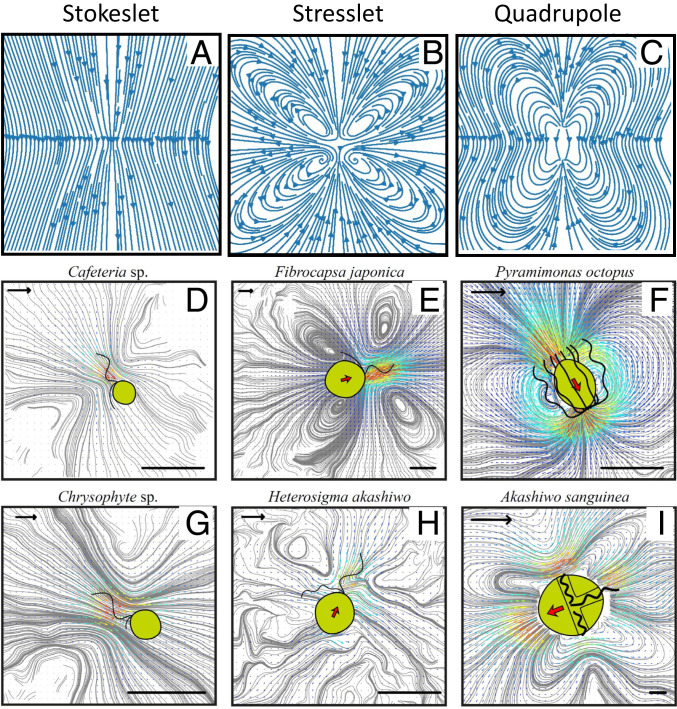
Fig. 3.
Streamlines of three types of point force models (A–C) and time-averaged flow fields around six flagellate species (laboratory frame) (D–I). In D and G, the cells are sessile, and the flow fields resemble a stokeslet (A). In E and H, the cells are free-swimming “pullers,” with the flagellar forces primarily at the front, and the flow fields resemble a stresslet (B). In F and I, the cells are free-swimming and have the forces in a mostly equatorial position. These produce flow fields that resemble various forms of quadrupoles (C). (Length scale bars: 10 μm; flow scale bars [arrows in upper left corner]: 100 μm s−1.)

