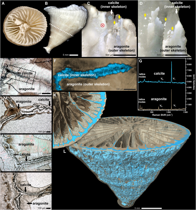
Fig. 1.
Extant specimen of the solitary scleractinian coral Paraconotrochus antarcticus with a two-component calcitic (inner)–aragonitic (outer) skeleton. Distal (A) and lateral (B) views of the calice are shown. (C and D) The growth edges of the septa and wall exhibit a calcitic inner skeleton (white arrows) overgrown by an aragonite (outer) skeleton (yellow arrows); blue- and red-crossed circles mark the position of micro-Raman analyses. (F) A Raman map (region marked in E) showing the distribution of calcite (blue) and aragonite (beige) in a skeleton sectioned transversely. (G) Raman spectra (from 0 to 1,500 cm−1 that include both lattice and internal [v1, v4], vibrational modes) of coral aragonite (beige) and calcite (blue) collected from regions indicated in C. (H–J) Transverse sections of adult (H), juvenile (I), and early juvenile (J) parts of the calice. Distinct boundaries (i.e., heteroepitaxy) between the crystal-transparent calcitic regions (with dark RADs) and the brownish aragonitic regions are visible. (K and L) X-ray computed tomography visualization of the calcitic inner (blue) and the aragonitic outer skeleton (semitransparent beige) up to the level indicated with a dashed line in B. (A–C, E, and G) ZPAL H.25/114; (D) ZPAL H.25/115; (E and F) ZPAL H.25/116; (H, I, and J) ZPAL H.25/117.

