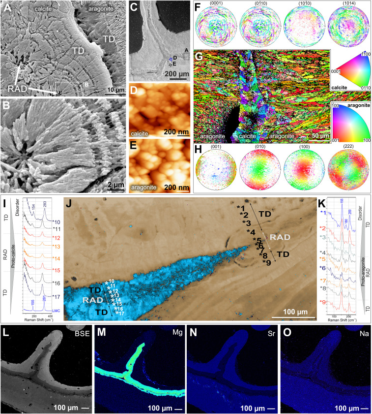
Fig. 2.
The microstructural, crystallographic, and geochemical features of calcitic and aragonitic regions of Paraconotrochus antarcticus skeleton. (A and B) The calcitic inner skeleton consists of RADs (arrows) and fibrous layers (i.e., TDs). (C–E) SEM of a transversely sectioned skeleton with a dashed frame indicating the region enlarged in A, whereas blue (D) and black (E) circles mark areas observed by AFM. Both calcitic (D) and aragonitic (E) skeletal parts have a nanogranular texture, typical of biominerals. (F–H) The sharp crystallographic boundary between the inner calcitic and outer aragonitic skeleton (G); in both regions, crystals have their a and b axes rotating around a c axis (turbostratic distribution: calcite (F) in the plane 104 and aragonite (H) in the plane 222. (I–K) The Raman spectra in RADs and neighboring TD regions (numbers in J mark measurement points) indicate disordered material in both calcite (I) and aragonite RADs (K), consistent with biogenic formation from amorphous precursors. (J) A Raman map. (L–O) Back-scattered electron (BSE) and electron microprobe images show the expected contrasting trace-element distributions, with calcite enriched in Mg (M) and depleted in Sr (N) and Na (O) compared with aragonite. (A–E and L–O) ZPAL H.25/117; (F–K) ZPAL H.25/116.

