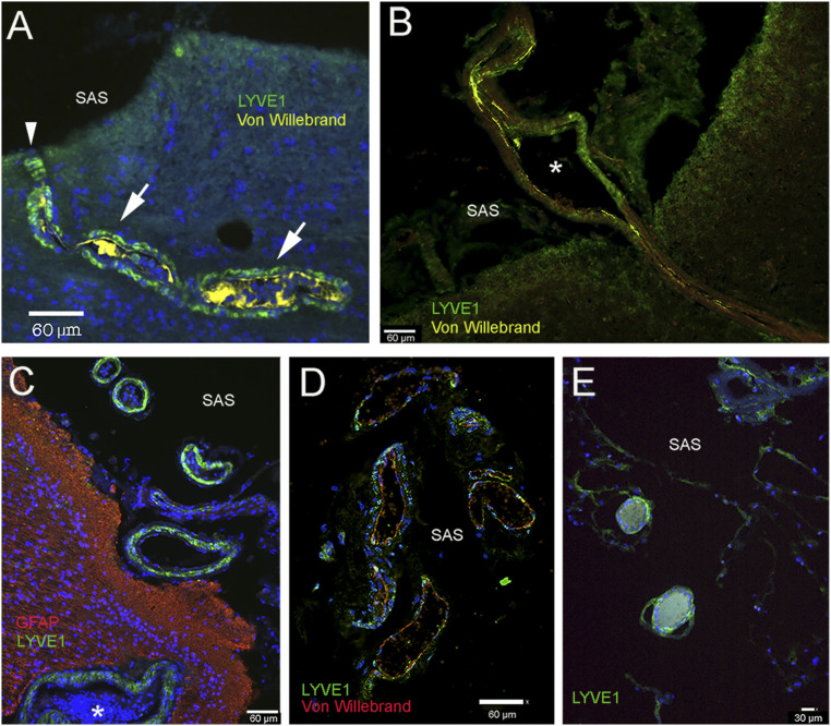
Fig. 3.
Lymphatic markers in the wall of vessels connecting with the subarachnoid space (SAS) over the frontal and parietal cortex. A–C and E are taken from patients with underlying neurological diseases, and C is from a patient with no neurological disease. A and B show vessels in the frontal cortex connecting with the subarachnoid space (SAS) in the arachnoid trabeculae. The outsides of the vessels are delineated by LYVE1-positive green cells, while the vascular endothelium is in yellow (staining von Willebrand factor). In A, the green LYVE1-positive cells that follow the vessel are labeled with arrows. The entry point of the vessel at the border of the SAS is labeled with an arrowhead. C–E show vessels in the SAS. In C, the outside wall of the vessel is LYVE1 positive (green), and processes of astrocytes (GFAP) at the surface of the brain are red. The asterisks in B and C label vascular lumina. (Scale bars: A–D, 60 μm; E, 30 μm.)

