Fig. 4.
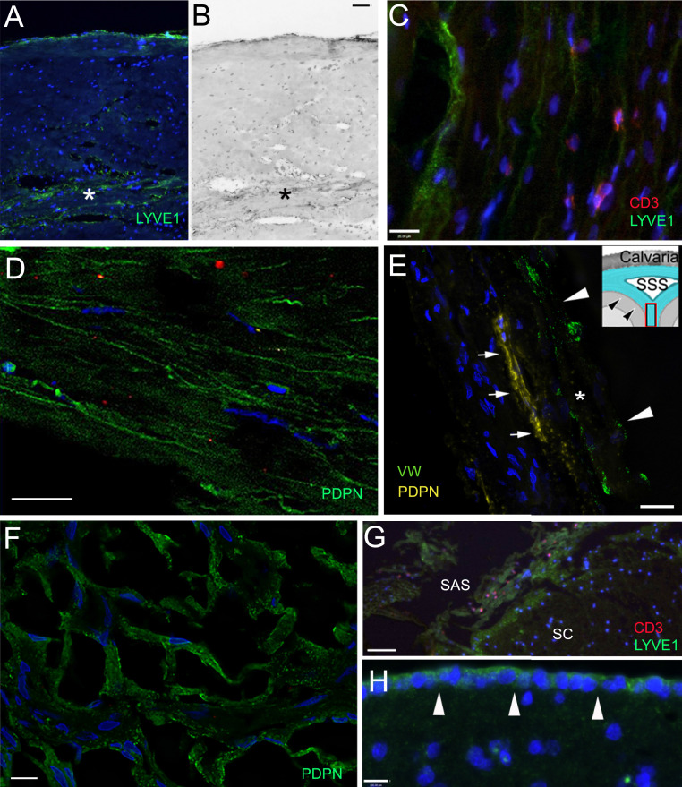
Lymphatic markers (LYVE1 and PDPN) in the meninges. Sections of the meninges from persons died of nonneurological causes (A, B, and E–G) and who suffered from neurological disease (C and D). (A–D) Upper (periosteal) and lower meningeal segments of the dura mater from the sagittal sinus; (E) falx cerebri; (F and G) arachnoid; (H) pia mater. (A and B) Bright-field image of the same field; in the dura mater LYVE1-positive lymphatic channels are stained green. The inner periosteal layer of the dura is adherent to the periosteum of the cranium. This layer is composed of tough compact collagenous fibers. The meningeal layer (labeled with a star) faces the arachnoid membrane. This layer is less compact; blood vessels and lymphatic elements fill up the intermembranous spaces. The capillaries and postcapillary venules are fenestrated (no BBB). (C) LYVE1-positive lymphatic cells and scattered T cells (stained in red with CD3 antibody) can be seen among the collagenous membranes of the dura. D shows podoplanin (PDPN; another accepted marker of lymphatic endothelial cells) positivity (green) in the dura mater in the inner surface of the basis of the cranium, in the cavernous sinus. E demonstrates a PDPN-positive (yellow fluorescence) lymphatic channel (small arrows) next to a sectioned vessel labeled with an asterisk and arrowheads (vascular endothelium is positive for von Willebrand factor; green fluorescence) in a thin section (taken from a Z stack at 1-µm optical thickness) of the falx cerebri (the anatomy is shown in the Inset, where arrowheads point at the dura mater and the square shows the approximate area of the falx cerebri where the sample was taken from. F demonstrates podoplanin staining of the arachnoid (from the Greek word for spider). It resembles a spider web. In G, many LYVE1-positive epithelial cells can be observed in the arachnoid covering the spinal cord (SC) and red fluorescent CD3-positive T cells are occasionally present near the LYVE1-positive connective tissue cells. (H) The cells of the pia mater (pointed at by white arrows) on the surface of the frontal cortex express LYVE1 (green). SAS, subarachnoid space; SSS, superior sagittal sinus. (Scale bars: A and B, 100 µm; C–F, as labeled.)

