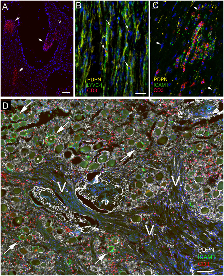
Fig. 8.
Expression of adhesion molecules in relation to T cells and lymphatic markers in the trigeminal nerve. All panels are from subjects with no known neurological disease. The trigeminal nerve was stained for CD3 (red), Lyve1 (green), and podoplanin (yellow). A shows a group of T cells at the sectioned surface of the nerve bundles (short arrows) suggesting an accumulation of T cells under the perineurium covering the nerve. One branch of the fifth cranial nerve is labeled with the roman numeral V. B demonstrates the colocalization of two lymphatic markers (podoplanin and Lyve1) in the endoneurial connective tissue and shows T cells (arrows) next to double-stained membranes. In C, yellow color represents PDPN, and the area where the red (CD3-positive) T cells gather is also strongly expressing the adhesion molecule ICAM1 (shown in green). A few PDPN-expressing cells with elongated nuclei are pointed at with arrows. ICAM1 seems to be expressed in the proximity of T cells. In D, the cross-section of the trigeminal ganglion and trigeminal branches from a strangulated victim is shown to demonstrate the T cell accumulation. Triple staining shows podoplanin (in white); ICAM1, the adhesion molecule known to be involved in T cell adhesion/migration (in green); and CD3 (in red) labeling all T cells. Large ganglion cells (some are labeled with stars) are clearly visible among the fibers. Note that—similarly to Fig. 7—only some of the ganglion cells are surrounded by ICAM1-positive green satellite cells (some of these are pointed out by arrows), while all other satellite cells are unstained for ICAM1 but still stained with PDPN (in yellow). (Scale bars: A and D, 100 µm; B and C, 50 µm.)

