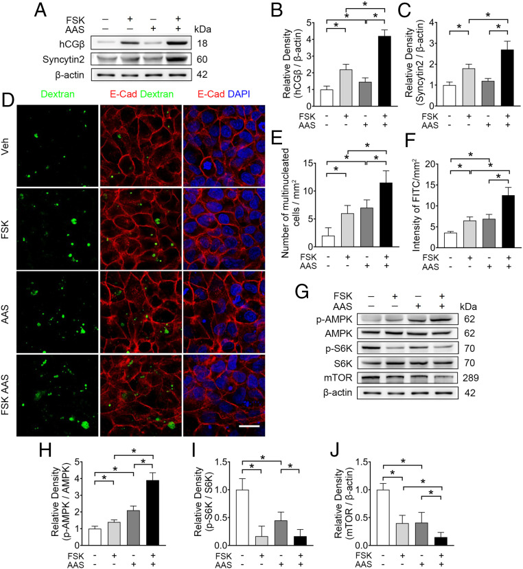
Fig. 2.
AAS simultaneously promotes syncytialization and macropinocytosis in BeWo cells. (A–C) Western blots (A) and corresponding quantification (B and C) of hCGβ and syncytin2 in BeWo cells cultured in AAS, with concentrations of Lys, Glu, and Arg at one-eighth of the normal levels with or without 20 μM FSK. (D) Representative immunostaining of dextran (green), E-cadherin (red), and DAPI (blue) in BeWo cells cultured in conditions detailed. (Scale bar, 40 μm.) (E and F) Quantification of multinucleated cell and FITC intensity. (G–J) Western blots (G) and corresponding quantification (H–J) of p-AMPK, AMPK, p-S6K, S6K, and mTOR in BeWo cells cultured in the AAS condition with or without 20 μM FSK. (H–J) Semiquantification of the relative density of p-AMPK/AMPK, p-S6K/S6K, and mTOR. Data were shown as mean ± SD and analyzed by one-way ANOVA test and Tukey–Kramer multiple comparison test based on at least three independent experiments. *P < 0.05.

