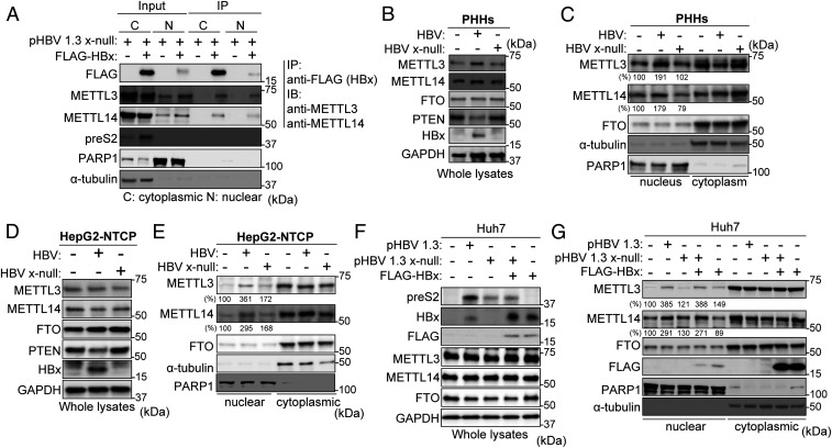
Fig. 4.
HBx protein interacts with METTL3 and 14 and stimulates their nuclear import. (A) Huh7 cells, which were transfected with pHBV 1.3 x-null, were treated with control or pcDNA 3.1 FLAG-HBx. Cytoplasm and nuclear lysates were immunoprecipitated with anti-FLAG, followed by immunoblotting for the indicated proteins. (B–E) PHHs were infected with HBV WT or x-null particles for 10 d (B and C). HepG2-NTCP cells were infected with the same amount of virus particles of HBV WT or x-null for 24 h and then further incubated for 9 d (D and F). The HBV-infected PHHs and HepG2-NTCP cells were harvested to assess immunoblotting from whole lysates (B and D) or isolated nuclear and cytoplasmic biochemical fractions (C and E). (F and G) Huh7 cells were transfected with pHBV 1.3 or HBV 1.3 x-null together with pcDNA3.1 FLAG-HBx or pcDNA3.1 (control). The indicated proteins were analyzed by immunoblotting (F). Immunoblot analysis of isolated nuclear and cytoplasmic biochemical fraction from indicated cells (G). In C, E, and G, the METTL3 and 14 expression levels relative to FTO from three independent experiments were quantified using ImageJ.

