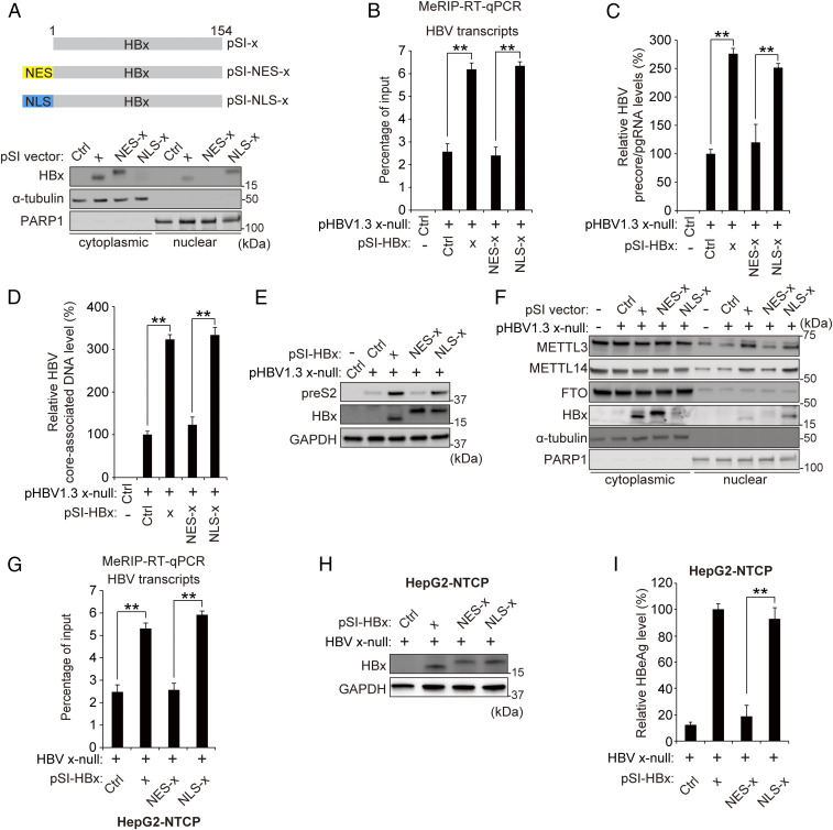
Fig. 5.
Nuclear localization of HBx is required for inducing m6A modification of viral transcripts. (A) Subcellular localization of HBx, NES-fused HBx, or NLS-fused HBx protein transiently expressed in Huh7 cells for 48 h was analyzed by immunoblotting. (B–E) Huh7 cells were transfected with HBV 1.3 x-null together with pSI-x, pSI-NES-x, or pSI-NLS-x for 48 h prior to quantification of MeRIP qRT-PCR (B), precore/pgRNA (C), core-associated DNA (D), or immunoblotting analysis for the indicated proteins (E). (F) Analysis of m6A methyltransferase levels in the cytoplasmic or nuclear fraction of HBV 1.3 x-null–expressed Huh7 cells, which were cotransfected with pSI-x, pSI-NES-x, or pSI-NLS-x. The indicated proteins were analyzed from cytoplasmic and nuclear lysates by immunoblotting. (G–I) HepG2-NTCP cells were infected with HBV WT or x-null particles. After 7 d, cells were transfected with pSI-x, pSI-NLS-x, or pSI-NES-x plasmid for 3 d. Cells were harvested to assess m6A methylation levels of HBV RNAs and PTEN mRNA (G) or for immunoblotting analysis for the indicated proteins (H) or HBeAg levels (I). In all panels, data are mean ± SD (**P < 0.01 by unpaired one-tailed Student’s t test).

