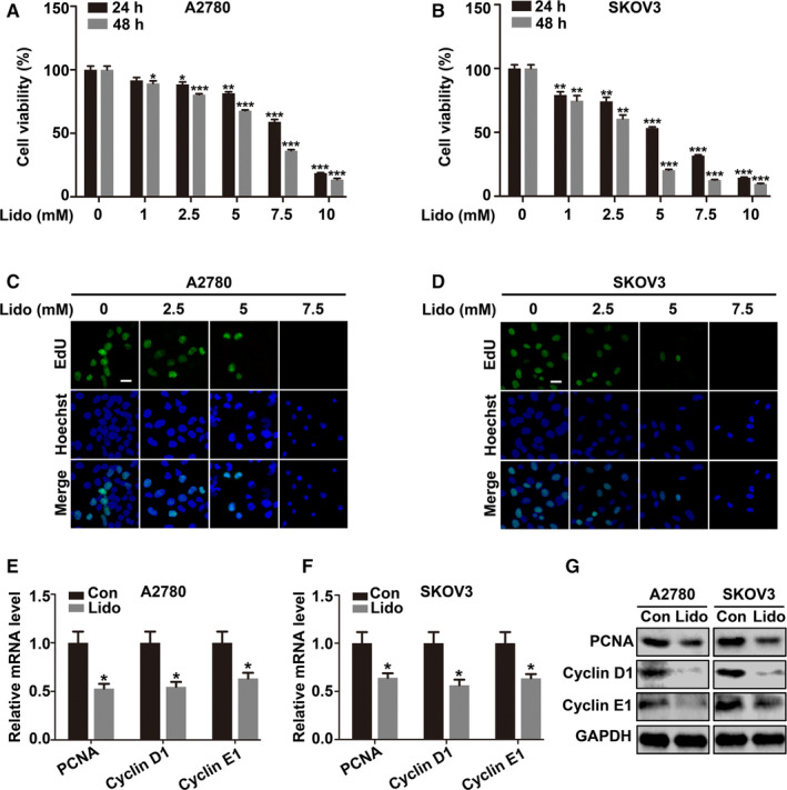
Figure 1.

Lidocaine inhibits the proliferation of ovarian cancer cells. A and B, A2780 and SKOV3 cells were treated with lidocaine (0, 1, 2.5, 5, 7.5, and 10 mM) for 24 and 48 hrs, respectively. CCK‐8 assay was used for cell viability evaluation. C and D, A2780 and SKOV3 cells were exposed to lidocaine (0, 2.5, 5, and 7.5 mM) for 48 hrs. Representative images of FITC‐labeled EdU (green) incorporation assay were presented. Hoechst 33342 (blue) was used for nuclei staining. Bar represents 50 μm. E and F, qRT‐PCR and (G) Western blot showed the mRNA and protein expression levels of PCNA, Cyclin D1, and Cyclin E1 in control and lidocaine‐ (5 mM) treated cells. GAPDH was used as an internal control. The data were presented as mean ±SEM (n = 9); *p < 0.05, **p < 0.01, ***p < 0.001
