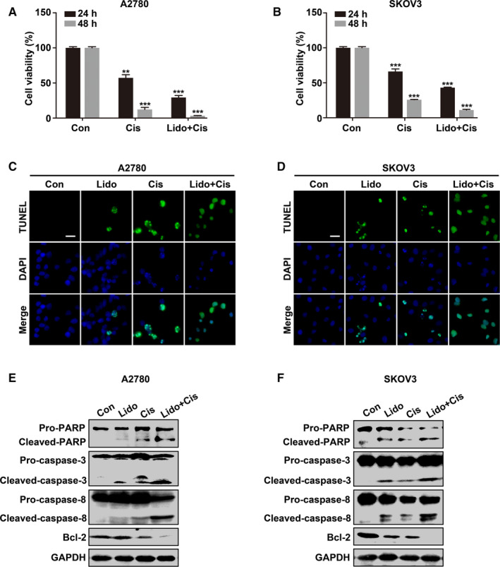
Figure 3.

Lidocaine sensitizes ovarian cancer cells to cisplatin in vitro. A and B, A2780 and SKOV3 cells were untreated, treated with cisplatin (10 μM), or cisplatin combined with lidocaine (5 mM) for 24 and 48 hrs, respectively. Cell viability was evaluated by CCK‐8 assay. C and D, Representative images of TUNEL (green)‐labeled apoptotic A2780 and SKOV3 cells. DAPI (blue) was used for nuclei staining. E and F, Western blot analysis of apoptotic marker proteins in control, lidocaine, cisplatin, cisplatin, and lidocaine combination groups. GAPDH was used as an internal control. Bar represents 20 μm. The data were presented as mean ±SEM (n = 9, n = 3); **p < 0.01, ***p < 0.001
