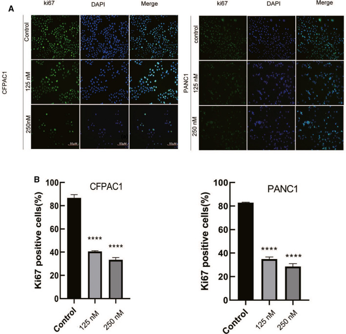
FIGURE 2.

Periplocin reduces the expression of Ki67 Immunofluorescence. A, After respectively treating CFPAC1 and PANC1 cells with 0,125, and 250 nm periplocin for 24 h, Ki67 Immunofluorescence showed cell proliferation at different periplocin concentrations. B, Ki67 Immunofluorescence and quantitative analysis. Scale bar, 50 μm. The results are expressed as the mean ±SD of independent experiments performed in triplicate. p ≤ 0.05 was considered to be statistically significant, * p < 0.05, ** p < 0.01, *** p < 0.001, **** p < 0.0001 versus the control
