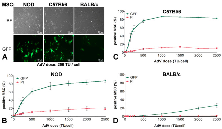
Figure 1.
Adenoviral transduction efficiency of murine mesenchymal stromal cells (MSC) isolated from three mouse strains. (A) Microscopy of non-obese diabetic NOD-MSC, C57BL/6-MSC, and BALB/c-MSC, transduced with 250 transduction units (TU)/cell adenovirus for GFP expression: BF—Bright field and GFP—fluorescence microscopy. (B–D) Dose-dependent transduction of MSC derived from NOD, C57BL/6, and BALB/c strains. The cells were incubated with increasing doses of adenovirus ranging from 0–2500 TU/cell; after 48 h the GFP expressing cells and the cell death determined by propidium iodide (PI) incorporation were evaluated by flow cytometry. The dose-dependent curves are different for NOD-MSC (B), C57BL/6-MSC (C), and BALB/c-MSC (D).

