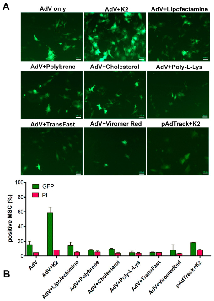
Figure 2.
Adenoviral transduction of murine mesenchymal stromal cells in the presence of different potential transduction boosters. To induce GFP expression, MSC were incubated with 250 TU/cell adenovirus alone (AdV only) or in the presence of the K2 Transfection System (K2TS) (AdV + K2), Lipofectamine 3000 (AdV + Lipofectamine), 10 μg/mL Polybrene (AdV + Polybrene), 2 μg/mL free cholesterol (AdV + Cholesterol), 1 μg/mL poly-L-Lysine (AdV + Poly-L-Lys), TransFast (AdV + TransFast), or Viromer Red (AdV + ViromerRed). In parallel, MSC were transfected with pAdTrack-CMV using K2TS (pAdTrack + K2). After 48 h, the GFP expression was observed by fluorescence microscopy (A), and the number of the GFP-expressing cells was determined by flow cytometry (B). The percentage of GFP-positive cells determined by flow cytometry is represented by green columns and that of the dead cells colored with propidium iodide (PI)—in red columns. As is revealed, the K2TS is the most efficient reagent for boosting the adenoviral transduction of MSC. Bars, 20 µm.

