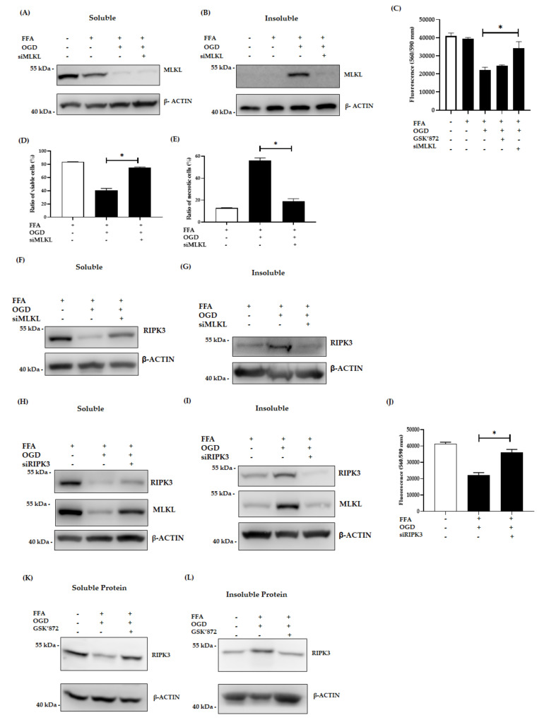
Figure 4.
Effect of siMLKL, siRIPK3 and GSK’872 treatment in FFA + OGD exposed AML-12 cells. (A,B): Soluble and insoluble MLKL protein expression after siMLKL treatment post OGD in FFA treated AML-12 cells respectively. (C): Cell viability was assessed by fluorometric quantitation. (D,E): Cells were stained with Annexin fluorescein and propidium iodide and analysed by flow cytometry for the ratio of percentage of viable cells and necrotic cells respectively. (F,G): soluble and insoluble RIPK3 protein expression siMLKL transfect FFA + OGD treated AML-12 cells respectively. (H,I): Soluble and insoluble RIPK3 and MLKL protein expression after siRIPK3 treatment post OGD in FFA treated cells respectively. (J) Cell viability was assessed by fluorometric quantitation. (K,L): Soluble and insoluble RIPK3 protein expression after 50 µm of GSK’872 treatment post OGD in FFA treated AML-12 cells respectively. β-ACTIN was used as the loading control for western blot. Data is represented as mean ± SD from 3 independent experiments. Comparison of cell viability in respective siMLKL and siRIPK3 transfected FFA + OGD treated groups and non-transfected FFA + OGD at * p < 0.0001. small interfering (si), mixed-lineage kinase domain-like pseudokinase (MLKL), receptor-interacting protein kinase 3 (RIPK3), Free fatty acid (FFA), oxygen glucose deprivation (OGD), alpha mouse liver 12 (AML-12) cell line, Beta-actin (β-ACTIN).

