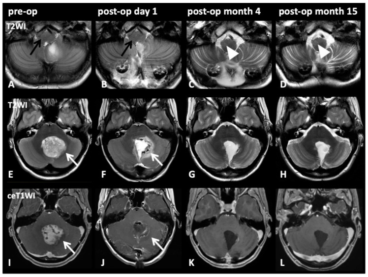
Figure 4.
Image series on patient number five from Table 2: Pre-op (1st column), post-op day 1 (2nd column), post-op month 3 (3rd column), post-op month 14 (4th column). T2WI axial slices on medullary level (A–D) and on pontine level (E–H) with corresponding pontine axial contrast enhanced T1WI (I–L). A 17-year-old female patient presented with cerebellar pilocytic astrocytoma affecting the left dentate nucleus (E,I; white arrows). Initially (A; black arrow) and after complete resection via a paravermal approach 6 days later (F,J; white arrows), no HOD is visible (B; black arrow). However, after 3 months (C, white arrow) and still after 14 months (D; cropped image, arrowheads), HOD with hyperintensity on T2WI on the right side can be found. No signs of tumor recurrence but only discrete scarring is visible after 3 and 14 months (K,L).

