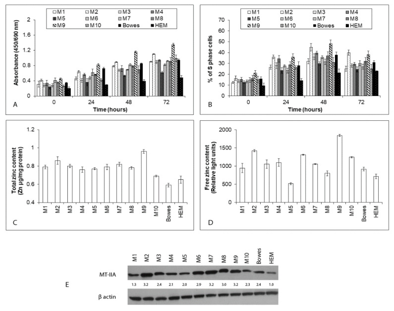
Figure 1.
The proliferation, zinc content, and metallothionein II A (MT-IIA) expression in human explant melanoma cells, Bowes cell line and normal human melanocytes HEM. Established explant human melanoma cultures (labeled M1–M10), human melanoma cell line Bowes and normal human melanocytes HEM were maintained upon standard laboratory conditions and their proliferation was determined by (A) colorimetric WST-1 assay measuring the cleavage of tetrazolium salt WST-1 by mitochondrial succinate dehydrogenases in viable cells and by (B) measuring the proportion of S-phase cells via EdU-specific fluorescence. Values represent means ± SD of at least three experiments. (C) Total zinc content in human explant melanoma cells (M1–M10), Bowes cell line and normal human melanocytes HEM. Zinc content was determined by absorption spectrometry. (D) Free (labile) zinc content in human explant melanoma cells (M1–M10), Bowes cell line and normal human melanocytes HEM as measured by microfluorometry of the zinc-specific dye Newport Green diacetate. Values represent means ± SD of at least three experiments. (E) The expression of metallothionein II-A (MT-IIA) in melanoma cell lysates as determined by immunoblotting analysis. The numbers in the blot image refer to fold increase or decrease in the density of the particular protein compared to the density of the same protein in HEM cells. Shown is one typical result of at least four experiments.

