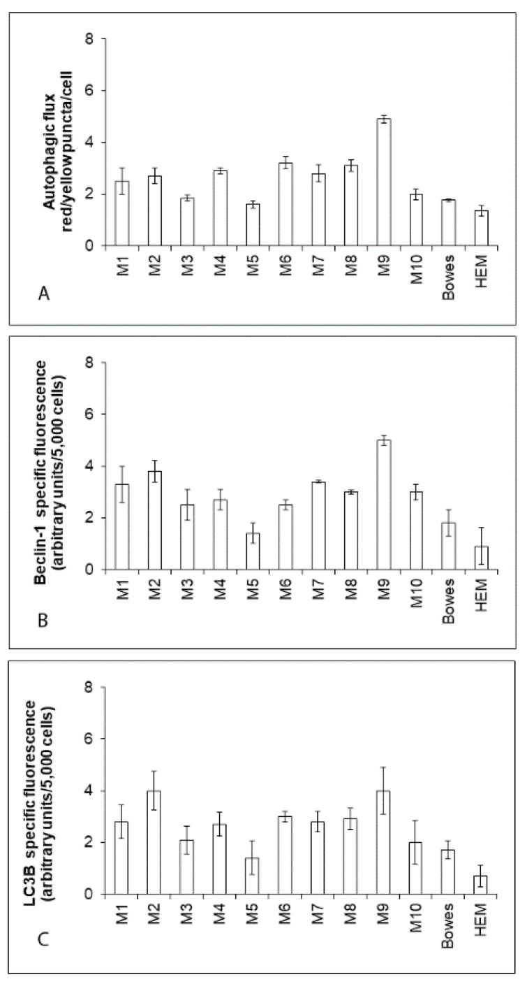
Figure 2.
Autophagic activity in human explant melanoma cells, Bowes cell line and normal human melanocytes HEM. (A) Autophagic flux (the rate of formation of autophagosomes and autophagolysosomes) in individual cells was determined with help of RFP-GFP-LC3B reporter system following cell transduction and subsequent quantitation of yellow (LC3B positive autophagosomes) and red (LC3B positive autophagolysosome) fluorescence with the determined ratio of yellow/red puncta per cell. (B,C) The expression of proteins Beclin-1 and LC3B in the examined cells were determined fluorimetrically. Values represent means ± SD of at least three experiments.

