Figure 3.
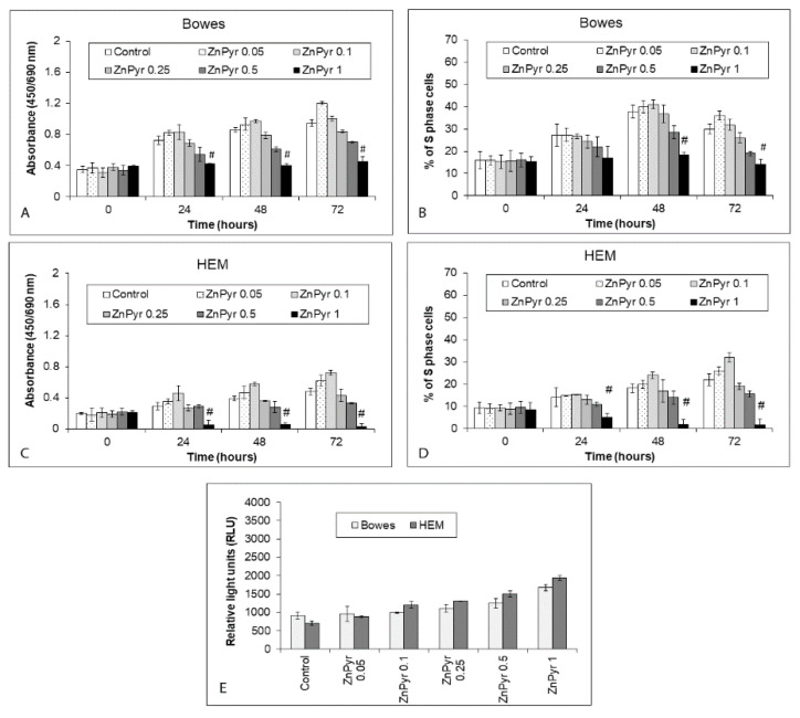
Effect of externally added zinc ionophore zinc pyrithione on the proliferation/viability and free zinc content of Bowes cell line and normal human melanocytes HEM during 72 h. Cells were exposed to zinc pyrithione in cultivation medium and (A–D) proliferation/viability was determined at individual time intervals with colorimetric WST-1 assay (measures the rate of metabolic conversion of tetrazolium salt WST-1 by mitochondrial succinate dehydrogenase in viable cells) and via measuring the proportion of S-phase cells using EdU-specific fluorescence. Values represent means ± SD of at least three experiments. # p < 0.05 Significantly lower than control at the same treatment interval with a one-way ANOVA test and Dunnett’s post-test for multiple comparisons. Free (labile) zinc content of treated cells (E) at 48 h was determined using fluorimetry of the zinc-specific dye Newport Green diacetate. Results represent means ± SD of five experiments.

