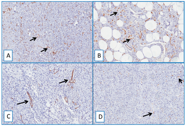
Figure 5.
Immunohistochemical analyses showed no detectable staining for WT1 in dermatofibroma (A), spindle cell lipoma (B), nodular fasciitis (C), and solitary fibrous tumor (D). Cytoplasmic staining of blood vessels served as a positive internal control (arrows) (immunoperoxidase; original magnifications 50× (A,D) and 150× (B,C)).

