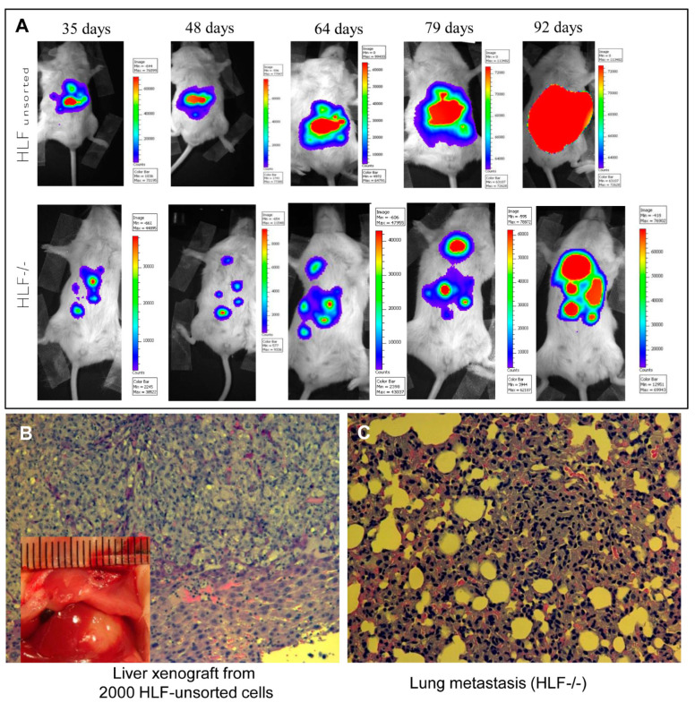
Figure 2.
In situ model of a liver xenograft and metastasis. Human hepatoma cells (HLF and HLF CD133−/EpCAM−) were injected directly into the medium lobe of the liver in NOD-SCID-IL2-γR(-/-) NSG mice under isoflurane anesthesia. These cells were transduced with lentiviral vector LUX-PGK-EGFP before the injection. One month after the injection, the mice were imaged with an IVIS 100 imaging system (Xenogen Corp.) every other week for longitudinal tracking of the formation of xenografts and metastasis over the next two months (A). (B) An in situ xenograft in the mouse liver was visible with histology confirmation. (C) Micrograph of the pulmonary metastatic invasion into alveoli or interstitial tissue (hematoxylin and eosin staining, 200×). The animal experiments were performed with the ethic approval by the institutional committee of animal care and use, and followed the NIH guidelines of experimental animals.

