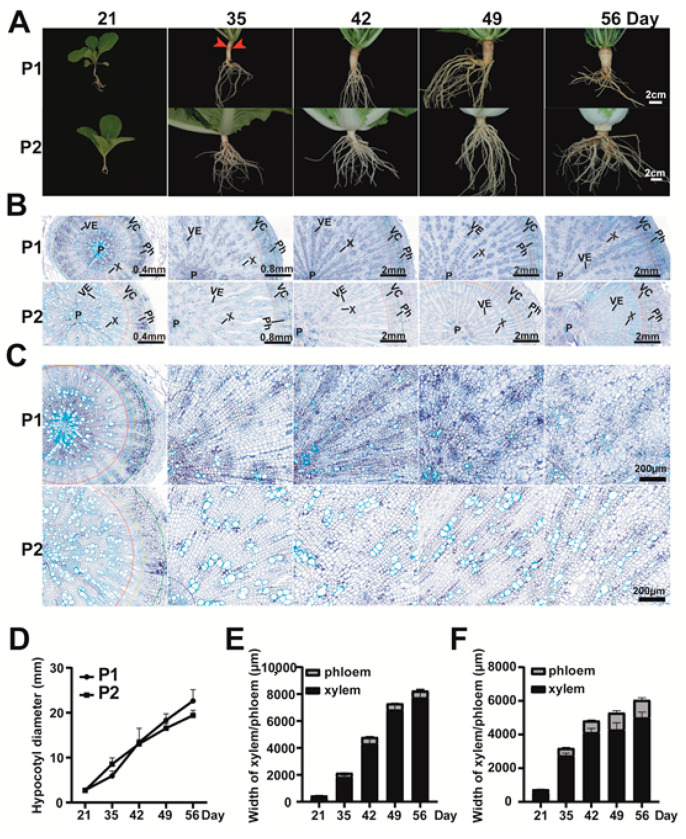
Figure 2.
Phenotypic and cytological observation of P1 and P2. (A) Phenotypic observation of the root and hypocotyl of P1 (turnip) and P2 (Chinese cabbage) at 21, 35, 42, 49, and 56 days after germination (DAG). Red arrows represent the cortex splitting phenotype. Three biological replicates were observed for each time point. Bar, 2 cm; (B) Bottom part of hypocotyls from P1 and P2 at 21, 35, 42, 49, and 54 DAG. P, pith; VE, vessel; X, xylem; Ph, phloem; VC, vascular cambium; (C) Xylem cells of P1 and P2 at 21, 35, 42, 49, and 54 DAG. Bar, 200 µm; (D) The hypocotyl diameter of P1 and P2 plants in different development stages; (E,F) The xylem width and phloem width of hypocotyls from P1 (E) and P2 (F).

