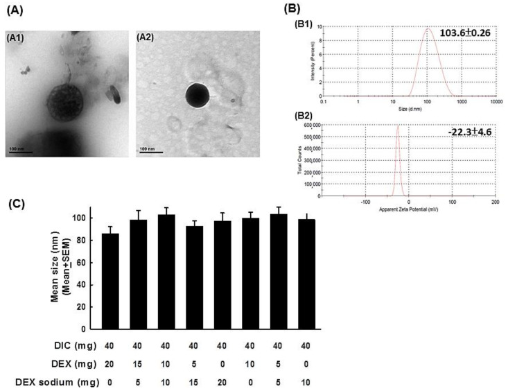
Figure 1.
Characteristics of various dosages of DIC/DEX-loaded liposomes. (A) Representative figures of DIC/DEX-loaded liposomal nanoparticles captured by transmittance electron microscopy. The left panel depicts an empty liposomal nanoparticle. The right panel depicts a liposomal nanoparticle that was loaded with DIC/DEX. (B) The representative figures of the size distribution and zeta potential in 40 mg/mL DIC combined with 5 mg/mL hydrophilic/hydrophobic DEX-loaded liposomal nanoparticles acquired by DLS analysis (B1: size distribution, B2: zeta potential). (C) Bar figures of size distribution of various dosages of DIC/DEX-loaded liposomal nanoparticles. Note: The average sizes of the DIC/DEX-loaded liposomal nanoparticles were approximately 200-400 nm. Abbreviations: DLS, dynamic light scattering; DIC, diclofenac; DEX, dexamethasone.

