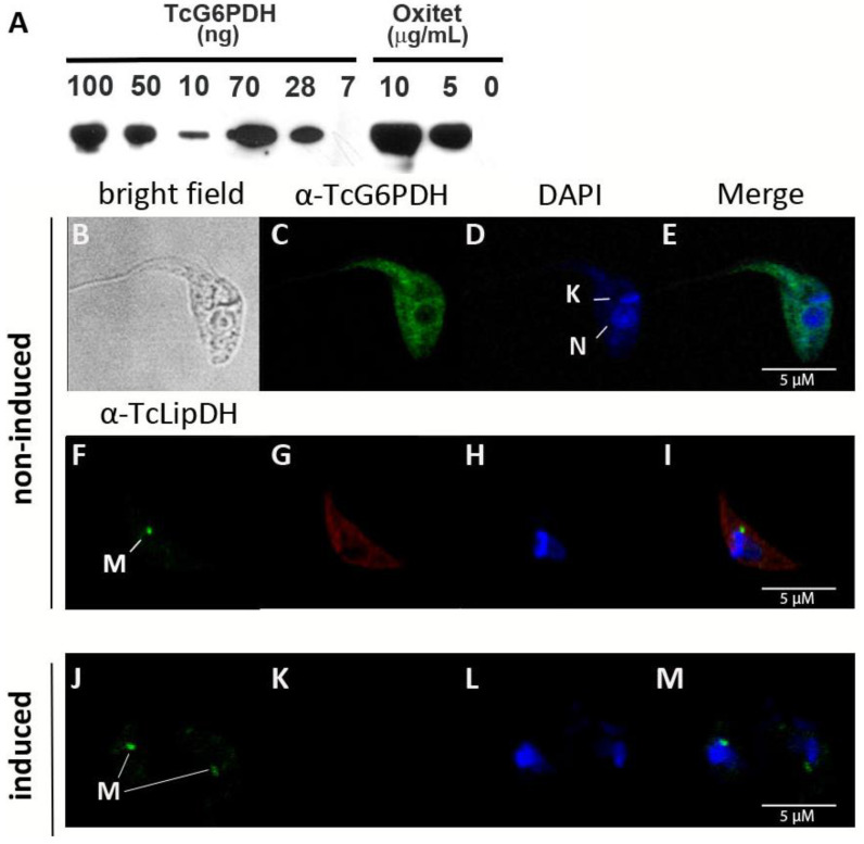
Figure 1.
Subcellular distribution of G6PDH in T. cruzi epimastigotes (strain Adriana). (A) Western blot analysis of different amounts of recombinant TcG6PDH (7, 10, 28, 50, 70 and 100 ng) and cell extracts of oxytetracycline induced (5 or 10 μg/mL: 1 × 10⁷ cells/lane) and non-induced (0 μg/mL: 1 × 10⁶ cells/lane) trypanosomes. Confocal microscopy of samples from induced (B–I) and non-induced (J–M) parasites. (C,G,K) Indirect immunofluorescence using anti-TcG6PDHL and secondary anti-mouse serum conjugated to Alexa 488 (green signal) or Alexa 594 (red signal), respectively. (D,H,L) Mitochondrial (K) and nuclear (N) DNA was stained with DAPI (blue signal). (F,J) signal corresponding to the mitochondrial lipoamide dehydrogenase (M) detected with specific anti-serum against the T. cruzi protein. (E,I,M) Merge images of the fluorescence staining.

