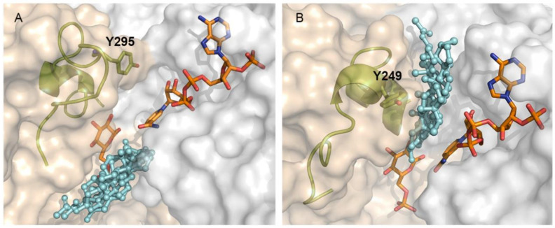
Figure 7.
Differential binding site of steroids derivatives to T. cruzi and human G6PDH. (A) Catalytic model of TcG6PDH (left side) and (B) HsG6PDH (right side), NADP+ and G6P are shown as orange sticks. Steroid derivatives (cyan balls and sticks). The element containing the tyrosine residue (Y295 and Y249 in Tc- and Hs-G6PDH) is shown as cartoons and olive colored sticks. The N-terminal Rossmann-fold and the C-terminal dimerization domain of TcG6PDH are shown as having their surfaces colored in light-gray and wheat color, respectively.

