Figure 1.
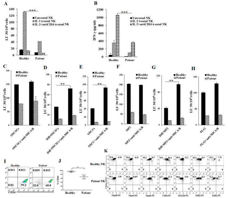
Cancer patients’ natural killer (NK) cells exhibit lower functional activity and NK cell-mediated antibody-dependent cell cytotoxicity (ADCC) when compared to healthy individuals’ NK cells. Purified NK cells (1 × 10⁶ cells/mL) from healthy individuals and pancreatic cancer patients were left untreated, treated with interleukin-2 (IL-2) (1000 U/mL), or treated with a combination of IL-2 (1000 U/mL) and anti-CD16 mAb (3 µg/mL) for 18 h and were added to 51Cr-labeled oral stem-like/poorly-differentiated oral squamous cancer stem cells (OSCSCs) at various effector-to-target ratios. NK cell-mediated cytotoxicity was measured using a standard 4-h 51Cr release assay against OSCSCs. The lytic units (LU) 30/106 cells were determined using the inverse number of NK cells required to lyse 30% of target cells × 100 (A) *** (p value < 0.001). NK cells were isolated and prepared as described in Figure 1A for 18 h before the supernatants were harvested and the levels of IFN-γ secretion were determined using single ELISA (B) *** (p value < 0.001). One of 20 experiments is shown in Figure 1A,B. Purified NK cells (1 × 10⁶ cells/mL) from healthy individuals and cancer patients were treated with IL-2 (1000 U/mL) for 18 h and were used as effectors in 51Cr release assay. OSCSCs and Mia PaCa-2 (MP2) tumors were differentiated as described in the Materials and Methods. OSCSCs (C), NK cell-differentiated OSCSCs (D), oral squamous carcinoma cells (OSCCs) (E), MP2 (F), NK cell-differentiated MP2 (G), and PL12 cells (H) were labeled with 51Cr for an hour, after which cells were washed to remove unbound 51Cr. Then, 51Cr-labeled tumor cells were left untreated or treated with anti-major histocompatibility complex-class I chain related proteins A and B (MICA/B) monoclonal antibodies (mAbs) (5 μg/mL) for 30 min. The unbounded antibodies were washed away, and the cytotoxicity against the tumor cells was determined using a standard 4-h 51Cr release assay. LU 30/106 cells were determined as described in Materials and Methods (C–H) ** (p value 0.001–0.01). The surface expression levels of CD16 of freshly purified NK cells from healthy individuals and cancer patients were analyzed using flow cytometry. IgG2 isotype antibodies were used as controls (n = 4) (I,J) ** (p value 0.001–0.01). Freshly purified NK cells from healthy individuals and cancer patients were analyzed for the surface expression levels of CD16, Nkp30, Nkp44, Nkp46, CD94, NKG2D, KIR2, and KIR3 using flow cytometry. IgG2 isotype antibodies were used as controls (K). One of eight representative experiments is shown in Figure 1K.

