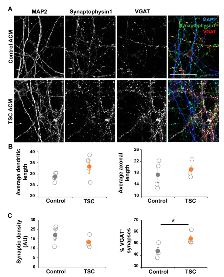
Figure 5.
Neurons in TSC ACM show synaptic changes. Control neurons were grown in ACM of either control or TSC astrocytes for 40 days. (A) Representative images of immunostaining at the end of the culture (day 57 of neuronal differentiation), showing dendrites by MAP2, all synapses by presynaptic marker Synaptophysin 1, and γ-aminobutyric acid (GABA) synapses by VGAT staining. (B) Morphological analysis showed no changes between TSC and control cells. Shown are the results of the average length of dendrites (MAP2+ neurites) and axons (SMI312+ neurites) per cell. (C) Synaptic analysis showed no significant changes in synaptic density, but the percentage of VGAT+ synapses was significantly increased in cultures with TSC ACM. ACM = astrocyte-conditioned medium, AU = arbitrary units. (A) Confocal images, scale bar = 50 μm (B,C) data points represent data for each iPSC line (average of two experiments) with solid data points representing the mean per condition ± SEM. * = p < 0.05 Statistical test: independent samples t-test.

