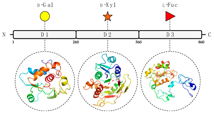
Figure 2.
Predicted GT domains of PBCV-1 encoded protein A111/114R. A111/114R domain analysis based on remote homology identified three putative GT domains labeled as D1, D2, and D3, located at the N-terminal (1 to 260 aa), central (261 to 559 aa), and C-terminal (560 to 860 aa) regions, respectively. Below individual domains are the corresponding three-dimensional protein models assigned by Phyre2 [18] based on alignments to known protein structures identified by their PDB entry: 1GA8 Chain A (D1), 2Z86 Chain D (D2), 2NZY Chain A (D3). Protein ribbon models are rendered using rainbow colors from N-terminus (blue) to C-terminus (red). The putative domain, predicted protein model, and sugar substrate are connected by the black dashes.

