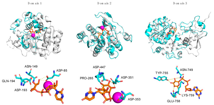
Figure 4.
Superpositions of A111/114R-D1, -D2, and -D3 with structural homologs. Individual domains of A111/114R (cyan) are shown independently as ribbon diagrams superimposed with known GTs (gray) bound to respective nucleotide sugars drawn as stick models (orange) and Mn2+ ion (magenta): N. meningitidis LgtC with UDP-fluorogalactose (PDB: 1GA8), E. coli K4CP with UDP-glucuronic acid (PDB: 2Z86), and H. pylori FucT with GDP-Fuc (PDB: 2NZY) are superposed with D1, D2, and D3, respectively (left to right). The corresponding active sites of D1, D2, and D3 are shown magnified below the complexed stereoviews with labeled residues proposed to be involved in sugar and ion coordination. Hydrogen bonds are represented as yellow dotted lines.

