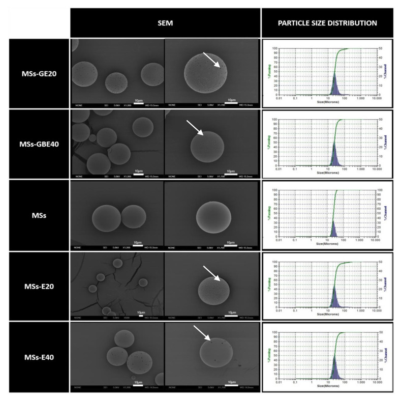
Figure 1.
Microsphere (MS) characterization. Morphological evaluation by scanning electron microscopy (SEM) and particle size distribution. Blank MSs (MSs); MSs/VitaminE(20) (MSs-E20); MSs/VitaminE(40) (MSs-E40) GDNF/VitE(20)-loaded PLGA MSs (MSs-GE20); GDNF/BDNF/VitE(40)-loaded PLGA microspheres (MSs-GBE40). SEM investigation showed the presence of spherical particles with comparable and regular size distributions, which were confirmed by particle size measurements. White arrows: pores on the MS surfaces. Scale bar: 10 µm.

