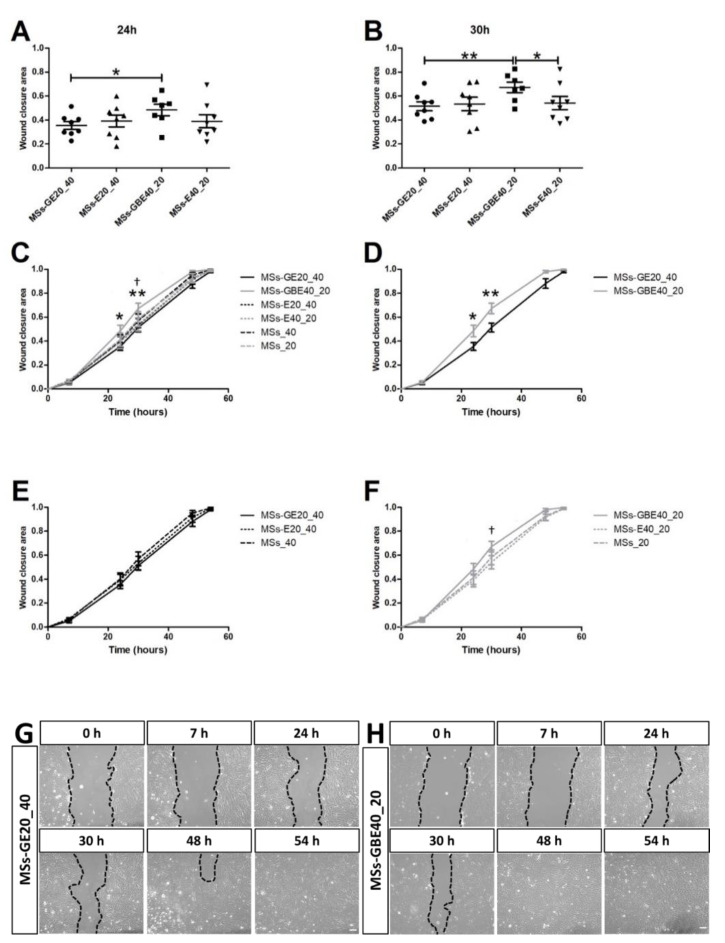
Figure 4.
Wound closure area in ARPE-19 cells. MSs-GBE (−) treated cells showed a more closed wound area than MSs-GE (−) treated cells both at 24 h (A) and 30 h (B) from scratch (p <0.05 and p < 0.01, respectively) and than MSs-E20_40 (- - -) at 30 h (B, p < 0.05). Graphs (C–F) and representative images (G,H) show a different pattern in timeline migration between MSs-GBE and MSs-GE treated groups in ARPE-19 cells at 0, 7, 24, 30, 48 and 54 h after scratching. Black dotted lines indicate the wound borders at the different time points and treatments. Blank MSs (MSs_20) and (MSs_40); GDNF/VitE(20)-loaded PLGA MSs (MSs-GE20_40); GDNF/BDNF/VitE(40)-loaded PLGA microspheres (MSs-GBE40_20). Scale bar: 100 µm. n = 6–8. * p < 0.05 and ** p < 0.01 MSs-GBE vs. MSs-GE; † p < 0.05 MSs-GBE vs. MSs-E40_20.

