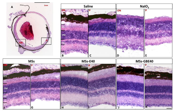
Figure 6.
Histology (hematoxylin and eosin staining) of retinas one week after intravitreal injection. (A) Whole eye section showing an optic nerve (ON) and peripheral (p) framed areas observed. Retinal section from eye injected with saline (B,C), sodium iodate (D,E), MSs (F,G), MSs-E40 (H,I), MSs-GBE40 (J,K). No alterations (swelling, vacuoles, missed cells) were observed in any studied group. Scale bar: 1 mm (A) and 100 µm (B–I). Blank MSs (MSs); MSs/VitaminE(40) (MSs-E40), GDNF/BDNF/VitE(40)-loaded PLGA microspheres (MSs-GBE40). Abbreviations: RPE: retinal pigment epithelium, OS: outer segments, ONL: outer nuclear layer, INL: inner nuclear layer, GCL: ganglion cell layer, ON: optic nerve, p: periphery.

