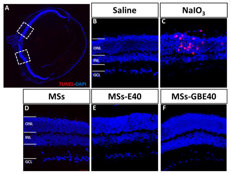
Figure 8.
TUNEL staining of retinal tissue. Representative micrographs of retina sections were evaluated for apoptosis by TUNEL assay at 1 week after intravitreal injection. (A) Whole eye section shows the retinal areas observed. (B) Retinal section from eye injected with saline without TUNEL positive cells. (C) Retinal section from eye injected with sodium iodate, a control positive of apoptosis. TUNEL-positive cells were identified with red fluorescence retinal section from eyes injected with MSs, MSs-E40 and MSs-GBE40 (D,E,F, respectively). TUNEL-positive cells were not found in eyes injected with PLGA and MSs. Nuclei of retinal cells were stained with DAPI (blue). Blank MSs (MSs); MSs/VitaminE(40) (MSs-E40), GDNF/BDNF/VitE(40)-loaded PLGA microspheres (MSs-GBE40). Abbreviations: ONL: outer nuclear layer, INL: inner nuclear layer, GCL: ganglion cell layer. Scale bar: 20 µm.

