Figure 1.
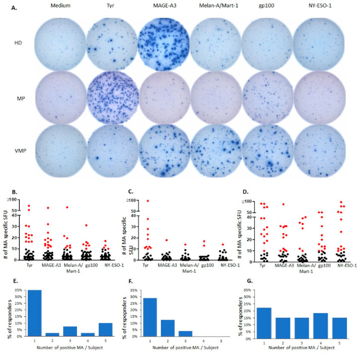
IFN-γ ELISPOT recall responses to melanocyte antigens. (A), Representative well images (96-well plate). The melanocyte antigens (MA) specified across the top were tested on PBMC of healthy donors (HD), untreated melanoma patients (MP), and MP vaccinated with AGI-101H (VMP). Well images are shown for a subject representative of each cohort. (B–D): The number of MA-specific CD8+ T cells in PBMC of 40 HD (B), 24 MP (C), and 27 VMP (D). For each subject, the IFN-γ ELISPOT recall response was tested after exposing the PBMC to the melanocyte antigens specified on the X-axis. Each data point represents the mean SFU count established in 250,000 PBMC/well, in triplicate wells. Positive T cell responses, as defined in Materials and Methods, are highlighted in red. (E–G): The number of MA eliciting positive T cell responses in 40 HD (E), 24 MP (F), and 27 VMP. PBMCs of each subject were tested in an IFN-γ ELISPOT assay for T cell reactivity to the five MA: Tyrosinase, MAGE-A3, Melan-A/Mart-1, gp100, and NY-ESO-1. The number of MA that elicited a positive response per donor (X-axis) is shown vs. the percentage of subjects in each cohort responding to that number of MA, specified on the Y-axis.

