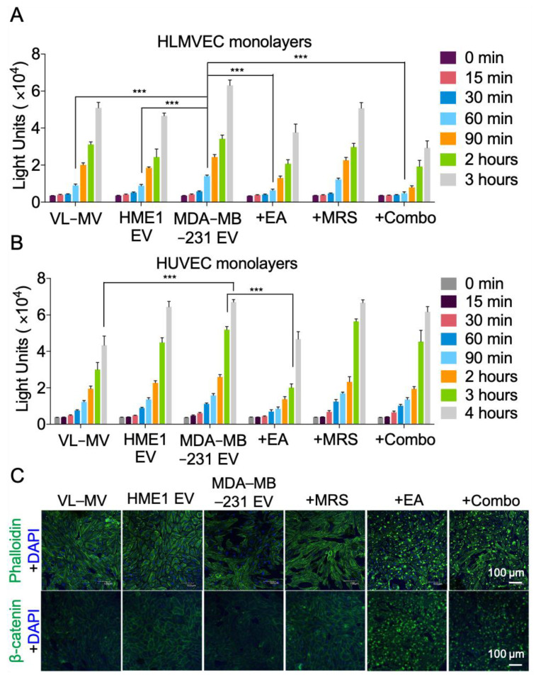
Figure 5.
NDPK inhibition ameliorates endothelial monolayer permeabilization by MDA-MB-231 EVs. (A) Permeabilization of HLMVEC and (B) HUVEC monolayers to FITC-dextran was measured using Matrigel-coated transwell chambers. Cells were treated with complete growth medium (VL-MV or VL-VEGF) with and without HME1 EVs, MDA-MB-231 EVs, and MDA-MB-231 EVs with MRS2179, EA, or a combination of both drugs. Intensity of FITC-dextran in the top chambers was measured at indicated time points as an indicator of enhanced permeability. n = 6. Mean ± S.E.M. *** p < 0.001 by one-way ANOVA and Tukey’s post-test. (C) CLSM images of HLMVEC monolayers immuno-stained for actin and β-catenin expression. All CLSM laser acquisitions were kept consistent, except for the bottom two images on the right, where 488 nm laser was enhanced to better visualize cells in these two treatment conditions. Cells were counterstained with DAPI. Scale: 100 μm.

