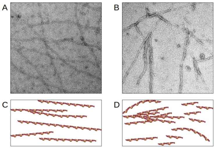
Figure 5.
Disruption of thin filaments induced by RCM cTnI-variants. (A,B) Electron microscopic images obtained with reconstituted cardiac thin filaments. (C,D) schematic presentation. (A,C) wild-type cTnI; (B,D) the RCM cTnI-variant p.R170W leads to shortened, wavy and partially aggregated thin filaments. The figure was created using PowerPoint (Microsoft).

