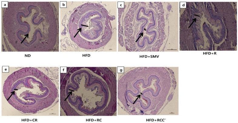
Figure 6.
Effect of plants extract on HFD-induced histopathological changes in H&E-stained aorta tissue. (a) Representative image of the liver tissue of normal control animal (ND). Animals fed with (b) HFD, (c) HFD + SMV, (d) HFD + R, (e) HFD + C, (f) HFD + RC, and (g) HFD + RCC’. Pathophysiological examination of the tissue sections was performed under a light microscopy at 200× magnification.

