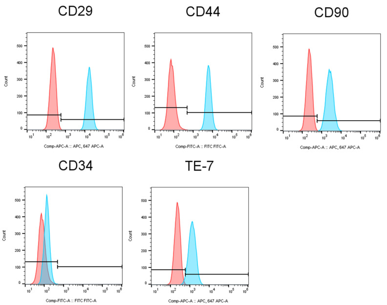
Figure 3.
Characterization of HGFs. The expression of stem cell markers (CD29, CD44 and CD90), hematopoietic stem cell marker (CD34) and fibroblasts marker (TE-7) on the surface of HGFs was analyzed by a BD FACSMelody™ Cell Sorter. The pink histograms indicate negative control, and the blue histograms show the stained cells, respectively.

