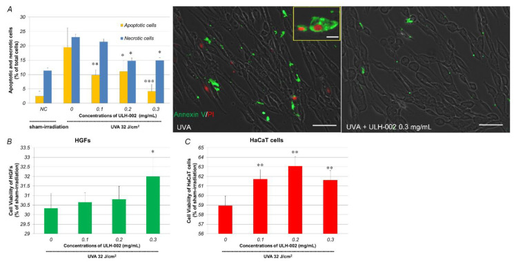
Figure 4.
Repressive effects of ULH-002 on UVA-induced cell damage in HGFs and HaCaT cells. (A,B), HGFs were pretreated with ULH-002 at different concentrations for 2 h. Cells were then irradiated with UVA (32 J/cm2). At 24 h after UVA irradiation, apoptotic/necrotic cells were detected by the Tali apoptosis kit. For the Tali analysis, cells were trypsinized and followed by Annexin V/PI staining. Cells expressing green fluorescence were counted as apoptosis. Cells expressed both green and red, or red only were counted as necrosis. For images, cells were washed twice with PBS(-) and then stained with Annexin V /PI. Green, Annexin V; red, PI. Scale bar indicates 25 µm in the broad images and 10 µm in the corner image (represents necrotic cells). NC, negative control group (sham irradiation). (B), at 48 h after UVA irradiation, cell viability of HGFs was measured by PrestoBlue assay. (C), HaCaT cells were pretreated with ULH-002 for 2 h and then irradiated with UVA. At 24 h after UVA irradiation, cell viability was measured by PrestoBlue assay. Data were expressed as mean ± SD. *, p < 0.05, **, p < 0.01, ***, p < 0.001 vs. “0”.

