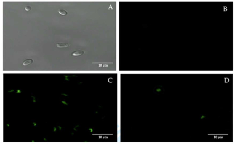
Figure 2.
Immunofluorescence assay using fluorescein isothiocyanate (FITC)-labeled anti-H. pylori IgG polyclonal antibodies. (A) bright field microscopy of C. albicans ATCC 90028 strain (negative control). (B) C. albicans ATCC 90028 strain (negative control) showing the absence of fluorescence. (C) H. pylori ATCC 43504 strain (positive control) showing fluorescence. (D) fluorescent intracellular H. pylori within yeast cells isolated from the vaginal discharge sample, cultured in Sabouraud agar supplemented with chloramphenicol, of a term pregnant woman. Micrograph D is a representative image of one of the triplicates of one of the vaginal discharge samples positive for the presence of intra-yeast H. pylori.

