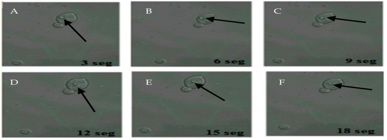
Figure 3.
Movement of H. pylori within yeasts of vaginal discharge origin. Confocal microscopy images taken 3 s apart using a Zeiss LSM780 NLO confocal microscope. (A–F) show the movement of H. pylori (arrows) inside the vacuole of a yeast cell. Light green color observed in the background represent remnants of fluorescein isothiocyanate (FITC). This figure is representative of images of one of the triplicates of one of the vaginal discharge samples positive for the presence of intra-yeast H. pylori.

