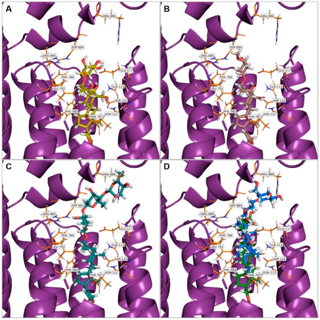
Figure 4.
Near view of cardiac glycosides (stick representations) docked into Na+/K+-ATPase (purple, image representations; orange, individual residues, stick representations) with the lowest binding energy mode. The cardiac glycoside binding site in the Na+/K+-ATPase is indicated. (A) Ouabain, (B) cymarin, (C) digitoxin, (D) hyrcanoside (in blue) and deglucohyrcanoside (in green). The images were taken using PyMOL 2.3.3 (Schrödinger, LLC, New York, NY, USA).

