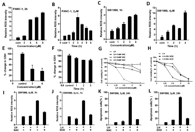
Figure 2.
LH induced-ROS accumulation is involved in its antitumor activity. (A), ROS levels in PANC-1 cells were assessed after 0, 2, 4, 6, 8 µM LH treated for 2 h by fluorescent probe DCFH/DA staining and determined by flow cytometry. (B), PANC-1 cells were incubation with 2 µM LH for 0, 1, 2, 3, 4, 5, 6 h, and ROS levels were measured as mentioned above. (C), SW1990 cells were treated with 0, 1, 2, 3, 4 µM of LH for 1 h and then ROS levels were assessed as mentioned above. (D), SW1990 cells were treated with 4 µM LH for 0, 1, 2, 3, 4 h, and then ROS levels were measured as mentioned above. (E), LH dose dependently decreased intracellular GSH levels. GSH levels were measured after SW1990 cells treated with 0, 3, 6 µM LH for 2 h by GSH and GSSG Assay Kit. (F), LH decreased intracellular GSH levels. GSH levels were determined after SW1990 cells disposed to LH at 4 µM for 0, 1, 2, 3 h. G-L, SW1990 cells were pretreated with NAC, SOD at indicated concentration for 30 min, the cell viability (G,H) was determined by CCK8 assay after incubated with 0, 1.25, 2.5, 5, 10 µM LH for 48 h, the levels of ROS (I,J) were determined by flow cytometry after incubated with 4 µM LH for 1 h, the apoptosis (K,L) was assessed by flow cytometry after incubated with 3 µM LH for 24 h.

